If your jaw lacks the height or density to hold an implant, a bone graft can create the stable foundation your new tooth needs. You may need a graft when prior tooth loss, gum disease, or long-term bone shrinkage leave too little support for an implant to fuse securely.
Understanding when grafting matters helps you avoid implant failure and unnecessary extra procedures later. It also frames the timeline you should expect for scans, grafting, and final implant placement. Working with an experienced oral surgeon in High Point, NC ensures this process is planned and executed with precision.
Expect clear explanations of graft types, who benefits most, recovery steps, and local factors in High Point, NC that can affect outcomes.
Table of Contents
ToggleUnderstanding Bone Health for Dental Implants
Healthy jawbone provides the mechanical support an implant needs and the biological environment for bone to fuse to the implant surface. You will need enough bone volume and blood supply at the exact implant site to achieve stable placement and long-term function.
The Role of Jawbone Density
Jawbone density determines how well an implant achieves primary stability at placement. Denser bone (commonly found in the front lower jaw) holds threads of the implant tightly, reducing micromotion during healing.
Less dense bone increases the risk of early implant mobility and may require a staged approach or wider/longer implants. Bone quality also affects the speed of osseointegration — the direct bone-to-implant connection.
Higher density can shorten the time before loading the implant. Low-density bone often needs extended healing or adjunctive measures like bone grafting or use of biomaterials with scaffolding properties.
Effects of Bone Loss on Oral Function
When you lose a tooth and the socket is left empty, the underlying alveolar bone begins to resorb. Resorption reduces ridge height and width, which can alter how nearby teeth contact and increase bite forces on remaining teeth.
Over months to years, facial support may diminish, changing lip and cheek contours. Functionally, insufficient bone can prevent placement of an implant in the ideal position for chewing and esthetics.
You may face compromised chewing efficiency and altered speech if multiple teeth are affected. There is also a greater risk of implant failure if a prosthesis places off-axis loads on marginal bone.
Assessment Techniques for Bone Volume
Clinicians combine clinical exam with imaging to measure bone volume and quality. Common tools include:
- Cone-beam CT (CBCT): provides 3D measurements of ridge width, height, and bone density at the planned implant angle.
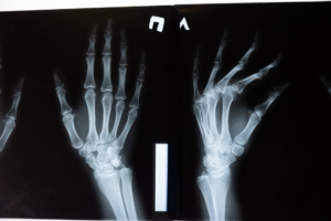
- Periapical and panoramic X-rays: useful for initial assessment of vertical height and adjacent anatomy.
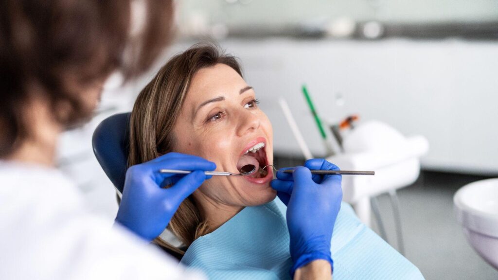
During the exam, your dentist will also probe soft-tissue thickness and evaluate previous extraction sites or pathology. Measurements focus on:
- Minimum ridge width for implant diameter
- Height above vital structures (sinus floor, inferior alveolar nerve)
- Cortical bone thickness and cancellous bone quality
If assessments show inadequate volume, the clinician will discuss graft options, timing, and projected healing intervals specific to your anatomy.
Types of Bone Grafting Procedures
You will choose a graft based on how much bone you need, where the defect sits in your jaw, and whether you prefer tissue from your body or a donor/synthetic source. Each option has trade-offs in healing time, surgical complexity, and long-term success.
Autografts Versus Allografts
Autografts use bone taken from your own body—commonly the chin, ramus (behind lower molars), or hip. Because the graft contains your living cells and natural growth factors, it integrates faster and carries the lowest risk of rejection.
Expect a second surgical site, added postoperative pain, and a slightly longer overall recovery. Allografts come from human donors processed and sterilized by tissue banks.
You avoid a second surgical site and most of the extra pain, but integration relies on the graft’s scaffold rather than living cells. Allografts are widely used in the posterior maxilla and narrow ridges; they have a high success rate when combined with proper implant timing and good oral hygiene.
Synthetic Bone Substitutes
Synthetic options include calcium phosphate, hydroxyapatite, and bioactive glass. These materials act as scaffolds that support new bone growth and gradually resorb over months to years.
You won’t face donor-site morbidity, and infection risk is low because products are sterile and consistent. Synthetic grafts suit small- to moderate-sized defects, sinus lifts, and guided bone regeneration when you want predictable material availability.
They sometimes integrate more slowly than autografts, so your surgeon may delay implant placement until radiographic evidence of sufficient bone density appears.
Socket Preservation Techniques
Socket preservation (ridge preservation) occurs right after a tooth extraction to limit bone loss and maintain ridge width for future implants. Your clinician places graft material—autograft, allograft, or synthetic—into the empty socket and covers it with a resorbable membrane or collagen plug.
This approach reduces the need for larger grafts later and preserves soft-tissue contours for esthetics. Healing timelines vary: small sockets may allow implant placement in 8–12 weeks, while larger or infected sites may require 4–6 months before safe implant placement.
Who Benefits Most from Bone Grafting
Bone grafting helps when your jaw lacks the height, width, or density to hold an implant securely. It rebuilds lost bone so an implant can be placed safely and last for years.
Patient Profiles with Significant Bone Loss
You benefit most when imaging shows less than 5–6 mm of bone height or thin ridge width under 4 mm. Age-related bone shrinkage, long-term denture wear, and trauma commonly cause these measurements.
If you have a narrow ridge, the implant diameter may exceed available bone, risking failure. A graft widens the ridge to allow standard implant sizes and proper implant positioning.
Medical history matters. Smoking, uncontrolled diabetes, and osteoporosis increase graft risk but don’t always rule you out.
Your surgeon will weigh these factors and may recommend specific graft types or medical management to improve success rates.
Impact of Gum Disease on Bone Structure
Advanced periodontitis destroys the supporting bone around teeth and creates uneven defects that compromise implant placement. If you have pocketing, loose teeth, or vertical bone loss on X-rays, grafting can rebuild sites after infection control.
You must complete periodontal therapy and show stable gum health before grafting. Treating infection first reduces graft failure risk and improves integration.
Grafts for periodontal defects differ from those after extractions. Your clinician may use bone substitutes plus growth factors to direct new bone where pockets once existed, restoring both volume and contour for implant esthetics and function.
Previous Tooth Extractions and Bone Resorption
After tooth loss, the socket walls collapse rapidly—most resorption occurs in the first 3–12 months. If you delayed implant placement beyond that window, you likely have horizontal and vertical bone loss that prevents ideal implant positioning.
Socket preservation at the time of extraction reduces the need for later grafting. If you didn’t have that, you may need block grafts, particulate grafts, or ridge augmentation to recreate volume.
Your treatment plan depends on defect size and location. Posterior molar sites often need more horizontal augmentation; anterior sites require precise contouring for esthetics.
Imaging guides whether a simple graft or staged reconstruction is necessary.
Preparation and Recovery Timeline
You will learn what checks happen before grafting, how long different grafts typically take to heal, and when your dentist will monitor progress and place the implant.
Pre-Procedure Evaluation Steps
Your dentist or oral surgeon will start with a focused exam and imaging. Expect a CBCT (3D) scan to measure bone height, width, and sinus proximity; these numbers determine whether you need onlay grafts, socket preservation, or a sinus lift.
Medical history review matters. Bring a list of medications, note any bleeding disorders, diabetes, or smoking—these change healing expectations and may require medical clearance or smoking cessation before surgery.
Lab tests and treatment planning usually follow. Your clinician may order bloodwork or coordinate with your physician for uncontrolled diabetes or anticoagulant management.
You’ll receive a written plan that states graft material (autograft, allograft, xenograft, or synthetic), anesthesia type, estimated costs, and an anticipated healing window so you can arrange time off and transportation.
Typical Recovery Periods
Healing time depends on graft type and location. For small socket preservation, expect about 8–12 weeks before adequate bone forms for implant placement.
Larger onlay or ridge augmentation often requires 4–6 months. Block grafts from your jaw or hip commonly need 5–6 months to integrate fully.
Sinus lifts follow a similar pattern: a lateral window sinus augmentation typically heals in 4–9 months, while less invasive transcrestal lifts may allow earlier implant placement.
Monitor pain and swelling: common symptoms peak 48–72 hours and then improve. Use prescribed antibiotics and NSAIDs as directed and avoid smoking or heavy exertion, which delay bone formation.
Follow-Up Appointments and Monitoring
Your first post-op check usually occurs 7–10 days after surgery to assess soft-tissue healing and remove sutures if needed. Subsequent visits at 4–8 weeks evaluate graft stability and soft-tissue health; your provider documents any signs of infection or graft exposure.
Radiographic checks matter. Expect a CBCT or periapical x-ray at the end of the healing window to confirm bone volume and density before implant planning.
Once imaging confirms adequate bone, your team schedules implant placement and reviews adjustments—antibiotic protocols, prosthetic timelines, and any additional grafting if needed.
Risks and Considerations for High Point, NC Residents
You should weigh specific surgical risks, anesthesia choices, and provider qualifications when planning bone grafting for implants. Costs, local infection rates, and recovery time in High Point can affect scheduling and follow-up care.
Potential Complications and Side Effects
Any surgery can cause pain, swelling, and bruising; bone grafting commonly produces these for several days to weeks. Infection at the graft site is the primary risk—your provider will prescribe antibiotics and give wound-care instructions to reduce that chance.
Graft failure or partial resorption is possible, particularly if you smoke, have uncontrolled diabetes, or have had prior radiation to the jaw. Nerve irritation or temporary numbness can occur when grafts are near sensory nerves; permanent nerve damage is rare but important to discuss before surgery.
If you plan implants soon after grafting, graft integration may delay implant placement. Expect follow-up visits and imaging to confirm bone growth; additional grafting may be required if integration is incomplete.
Local Anesthesia Versus Sedation Options
Local anesthesia alone numbs the surgical site and is appropriate for many small grafts; you remain awake and recover quickly. Local plus oral sedation (pill sedative) reduces anxiety while keeping you responsive; arrange a ride home after this option.
IV sedation provides deeper relaxation or a near-unconscious state and suits patients with severe anxiety, extensive grafts, or complex simultaneous implant placement. IV sedation increases monitoring needs and procedure cost, and you’ll need someone to drive you home and stay with you for several hours.
Discuss medical history, current medications, and any sleep apnea or heart/lung problems with the clinician. They will match anesthesia level to the procedure complexity and your health, while explaining fasting rules and recovery expectations.
Choosing a Qualified Dental Specialist
Look for a board-certified oral and maxillofacial surgeon or a periodontist with documented experience in bone grafting and implant placement. Check credentials, local patient reviews, and before-and-after cases specific to High Point practices to judge technical skill and outcomes.
Confirm the practice uses sterile technique, 3D imaging (CBCT), and contemporary graft materials. Ask how many grafts they place per year, complication rates, and whether they handle complications in-office or refer out.
Verify insurance or financing options, post-op support availability, and clear communication about timelines for graft healing and implant placement to avoid surprises.
Long-Term Outlook After Grafting
Grafted bone commonly supports implants for decades when the graft integrates well and you follow care recommendations. Predictable healing timelines and routine maintenance are the two factors that most influence long-term stability.
Expected Implant Success Rates
You can expect implant success rates after grafting to be similar to implants placed in native bone when the graft fully integrates. Published data and clinical practice typically report success rates in the 90–98% range at 5–10 years for properly placed implants after successful grafting.
Success depends on graft type (autograft, allograft, xenograft, or synthetic), surgical technique, and whether the graft achieved sufficient volume and density before implant placement. Smokers, uncontrolled diabetes, and poor oral hygiene increase your risk of complications and lower long-term success.
Your surgeon will assess bone quality with imaging and may delay implant placement until 3–9 months after grafting to maximize integration. If the graft partially resorbs, additional augmentation or alternative implant designs may be required to maintain function.
Ongoing Care for Bone Health
You must maintain excellent oral hygiene and attend regular dental visits to protect the grafted site and implant.
Brush twice daily with a soft brush. Floss or use interdental cleaners around the implant, and have professional cleanings every 3–6 months as recommended by your dentist.
Lifestyle and medical management also matter.
Quit smoking. Control blood sugar if you have diabetes.
Ensure adequate calcium and vitamin D levels.
Inform your dental team about medications (bisphosphonates, anticoagulants) that affect bone healing.
Periodic radiographs will monitor bone level and detect early bone loss so your clinician can intervene before major problems develop.